Tearing Problems
Symptomatic tearing is one of the most frustrating problems for patients. When tearing becomes excessive it can interfere with your vision, cause eyelid redness and irritation or be associated with an infection. Among golfers, tearing can add strokes to your game!
Tearing may be protective, as when dust blows into your eye, or may be responsive to dryness in the eye when the air-conditioning vent blows into your face. However, when tearing becomes a daily occurence or is associated with an infection, the cause should be investigated by a qualified physician.
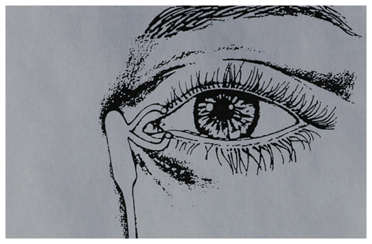
Initially, the eyelids should be evaluated due to their important role in tear distribution, drainage and globe protection. The eyelids should have an adequate punctal (tear duct) opening, a quick return snap and should close properly. The tear film should be inspected to ensure that an adequate amount and sufficient quality of tears are produced. The eyeball itself should be evaluated to ensure that an underlying intra-ocular problem is not causing secondary tearing.
The lacrimal (tear) duct can be evaluated with a dye test as an office procedure to determine whether the nature of the problem is due to a functional or complete obstruction of the duct. The dye test uses a vegetable dye (fluorescein) that flows with the tears to determine whether the tears enter the nose within an adequate period of time. If the dye fails to enter the nose the duct can be flushed with saline to determine whether (or not) it is open to irrigation. The results of the dye test determines how to proceed in treating the tearing problem.
The treatment of tearing related to underlying dryness may be as simple as increasing the use of ocular lubricants, such as artificial tears and ointments. The lacrimal puncta (tear duct openings) may be artificially plugged or permanently occluded to increase the amount of natural tears around the eye. Finally, the eyelid position may be altered to reduce the amount of ocular exposure.
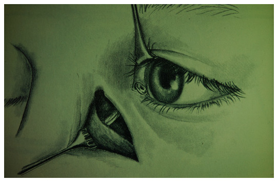
Tearing due to a true obstruction of the bony lacrimal duct requires surgery to restore normal drainage. The surgery involves creating a new passageway to bypass the obstruction. The tears continue to drain into the nose, but in a higher position within the nose. A temporary silicone (plastic) tube is used to maintain the opening during the healing phase. In rare cases, a permanent glass tube is used to completely bypass a severely damaged lacrimal drainage system. Tear drainage bypass surgery is typically done as an out-patient procedure under local anesthesia with sedation, or general anesthesia, as desired. There are typical three post-operative visits after surgery, the day after to check your vision, the week after to take out sutures and flush the new tear drainage channel, and the month after to ensure tear drainage and remove the temporary silicone tube.


